Functional Genomics Core

RRID:SCR_023433

Custom laboratory automation - Biomek FX

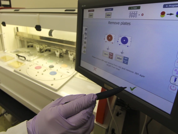
Our team specializes in writing custom programs (methods) and pipelines on our Biomek robots. Whether you need an entirely custom screening pipeline, or just the same few transfers done to a single plate week after week, we are happy to set up your project on our systems. It is our goal to train campus researchers to use our robots unassisted and on their own schedules. We make these instruments available to all trained users for independent use.

We have developed several programs (methods) that are designed to perform common cellular biology tasks in 96 or 384-well plates. These tasks include media swaps, PBS washes, reagent additions, etc. using the Biomek’s 96-tip pipetting head. These methods have been written with button interfaces that allow the operator to select from different plate types, numbers of plates, pipetting speeds and styles, and many other options to allow the operator to customize their usage independently.

We have also created programs to enable the operator to independently daughter and dilute compound libraries, perform cell based immunofluorescent assays (fix, permeabilize, probe with primary and secondary antibodies, wash, and stain).


Facility Email: fgc@arizona.edu
Image
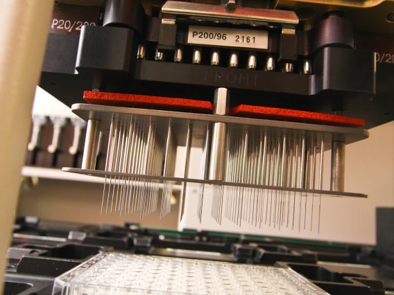
Biomek Pintool
Service
Location

Compound Screening

The Functional Genomics Core (FGC) works with investigators to perform all necessary assay optimization and validation. The FGC staff will translate your protocol into a high-throughput workflow on our liquid handling robots. Our team can perform your compound screen, work with your group to perform the screen, or train you to work independently on our systems. The primary limit on the throughput of the screen is the speed of the assay. Simple fluorescence based assays can be read on our CLARIOstar Plus microplate reader, while complex cell phenotyping can be performed on our Operetta CLS, binding assays can be run on our Octet RED384 BioLayer Interferometry (BLI) instrument.

The FGC is integrated with the Arizona Center for Drug Discovery (ACDD) providing resources for investigators conducting research in molecular biology to run live cell, biochemical and other assays. The ACDD, housed in the University of Arizona College of Pharmacy, creates the necessary infrastructure to bridge UArizona researchers with pharmaceutical partners to establish a collaborative environment to advance novel disease intervention findings beyond the laboratory setting. The ACDD works with scientists at the UArizona to develop and perform research projects in drug discovery. The ACDD also maintains compound libraries and provides assay design and optimization support.

For information or support on compound screening or drug discovery contact  Christopher Penton (pentoncm@arizona.edu) with the Arizona Center for Drug Discovery
Image
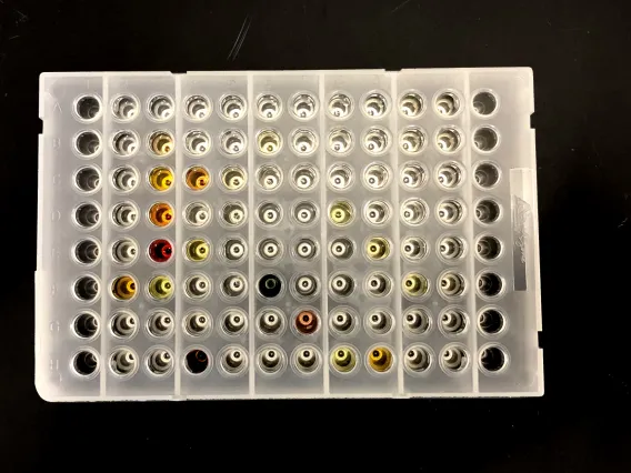
Abgene 96-well plate of compounds
Service
Location

Assay Consultation

The staff of the Functional Genomics Core will work with you to optimize your assay to produce the highest quality results on our high-throughput pipelines. We will help you choose the appropriate detection system for your experiment and validate the assay prior to high-throughput processing.


A Simple Statistical Parameter for Use in Evaluation and Validation of High Throughput Screening Assays

  • In this publication, a screening window coefficient, called “Z- factor,” is defined. This coefficient is reflective of both the assay signal dynamic range and the data variation associated with the signal measurements, and therefore is suitable for assay quality assessment.


Facility Email: fgc@arizona.edu

Image
CLARIOstar 384-well data
Service
Location

Singer RoToR HDA - pinning array robot

The Singer RoToR HDA pinning robot is a dedicated system for pinning arrays of yeast and bacteria. It is designed for rapid replication, arraying, mating and breakdown using 96, 384, 1536, or 6144 density of yeast, bacteria, or fungal libraries from one or more source plates to one or more destination plates. Transfers can be made from agar to agar, liquid to liquid, liquid to agar, or agar to liquid.

A Singer Rotor pinning robot is available for pinning arrays of yeast or other microbes. This instrument can be used to cross thousands of yeast strains to aid in yeast two hybrid screening, creation of novel yeast libraries, or replication of bacterial libraries. The Singer can also be use in conjunction with our Our Biomek FX liquid handling robots to create more complex pipelines, including re-arraying, cherry picking, and normalization/dilution of liquid cultures.

Our core has access to several custom yeast libraries or we can help you build you own.


Facility Email: fgc@arizona.edu
Image
Singer microbial arraying robot
Equipment
Location

Beckman Coulter Biomek FX systems - liquid handling robots

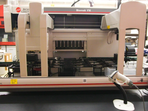
The FGC has two Dual arm Biomek FX liquid handling automation systems. The primary system is equipped with both a Liconic STX-44 live cell incubator and a large capacity Cytomat ambient storage hotel. The Liconic STX-44 incubator has temperature control, humidity control, CO2 control and can shake up to 1200 RPM. This incubator is capable of meeting the needs of most cell types for live cell screening. This system is also equipped with a detachable compound pinning tool, sonicating wash station, and pin drying fan. This robot can perform precision pipetting down to 0.2 µl and precision pinning down to 10 nl.

The second Dual arm Biomek FX liquid handling automation system is equipped with both a large capacity incubated Cytomat storage hotel and and a large capacity Cytomat ambient storage hotel. This system is configured to perform a wide array of liquid handling operations. Both systems can perform high-throughput cherry-picking and plate refactoring, dilution and normalization, and the automation of most molecular biology protocols.


Facility Email: fgc@arizona.edu
Image
Biomek robot with hotels
Equipment
Location

Operetta CLS - high-content analysis microscope imaging system

The Operetta CLS high-content analysis system has all the speed, sensitivity and resolution you need to characterize fine sub-cellular details . The Operetta is designed for fully-automated high-capacity, quantitative cellular analysis of fixed or live cells. It contains the highly sensitive sCMOS camera that provides a large field of view and high resolution image capture. The Operetta's Harmony software lets you find even subtle phenotypic changes.
Features:

  •     Objectives
    • 5x Air Objective: NA 0.16, WD 12.1 mm (field of view: approx. 2583 μm x 2583 μm)
    • 10x Air Objective: NA 0.3, WD 5.2 mm (field of view: approx. 1292 μm x1292 μm)
    • 20x Air Objective: NA 0.4, WD 8.28 mm (field of view: approx. 646 μm x 646 μm)
    • 20x Water Immersion Objective: NA 1.0, WD 1.7 mm (field of view: approx. 646 μm x 646 μm)
    • 40x Water Immersion Objective: NA 1.1, WD 0.62 mm (field of view: approx. 323 μm x 323 μm)
    • 63x Water Immersion Objective: NA 1.15, WD 0.6 mm (field of view: approx. 205 μm x 205 μm)
  • 4.7Mpx sCMOS camera (2160x2160), 16 bit resolution, 6.5um pixel size
  • Motorized 6 position objective turret with 3 air and 3 water immersion objectives
  • High resolution, linear drive scanning stage
  • High speed precision laser autofocus
  • Transmitted light unit (LED: 740nm) for Brightfield and Digital Phase Contrast imaging
  • Emission filters:
    • 430-500 nm
    • 470-515 nm
    • 500-550 nm
    • 525-580 nm
    • 570-650 nm
    • 655-760 nm
  • Spinning Disk enables optical sectioning
  • Confocal imaging improves optical resolution and contrast
  • Pinhole disk reduces out-of-focus light
  • LED light engine equipped with 8 high power LEDs with the following excitation wavelengths:
    • 355-385 nm
    • 390-420 nm
    • 435-460 nm
    • 460-490 nm
    • 490-515 nm
    • 530-560 nm
    • 615-645 nm
    • 650-675 nm
  • Environmental Controls for live cell experiments:
    • Temperature can be controlled between 37°C - 42°C
    • CO2 concentration can be controlled between 1-10%

Tips & tools for data collection and interpretation:

Revvity’s Harmony software is used to acquire images on the Operetta CLS high-content fluorescent microscope and can also be used to create custom automated image analysis methods.

Revvity has an interactive fluorescence modeling tool  that enables you to select the excitation LEDs and your possible emission spectral wavelengths to help construct an effective assay.  

Harmony, like most automated image analysis software, requires the use of fluorescent dyes, antibodies, or expressed proteins to tag the nucleus and/or the cytoplasm to differentiate separate regions of interest from background. To use "image segmentation" to identify cells and cell compartments, fluorescent channels are a critical starting point for all analysis methods to ensure successful image acquisition, and to allow for the creation of robust data analysis methods. Brightfield cannot be used to perform image segmentation within the Harmony software.
Recommended dyes for fixed cell assays:

  • Hoechst: DNA/Nuclei stain (available in the FGC)
    • Ex/Em: 361/497 nm
  • Phalloidin: Cytoskeleton/ ECM stain (available in the FGC)
    • Available in multiple fluorophores (350, 405, 555, and 647)
  •  DAPI (4′,6-diamidino-2-phenylindole): DNA/Nuclei stain
    • Ex/Em: 350/470 nm

Recommended dyes for live cell assays:

Additional resources for assay design:

Issues with culturing cells in 96-well plates:


Facility Email: fgc@arizona.edu

Image
Operetta HCS
Equipment
Location

Octet RED384 - protein interaction detection system

Campus users of the Biacore T200 SPR system should consider using BLI as an alternative.

The Octet RED384 is a non-fluorescence-based, high-throughput, bench top, protein interaction detection system that uses Bio-Layer Interferometry (BLI) technology, and can analyze up to 16 samples in parallel for affinity, kinetics or active protein concentration determination from samples in 96- or 384-well microplates.  It is a powerful detection platform for kinetic measurements & quantitation using the widest array of label-free binding assays available including ready made, custom coated, Biosensors:

    Streptavidin
    Protein A
    Protein G
    Protein L
    Ni-NTA
    Anti Penta-Histidine
    Anti-GST
    Anti-Human-Fab(CH1)
    Amine Reactive (NHS-EDC)
    Amino-Propyl-Silane
    Anti-Mouse
    Anti-Human

The closest technology to BioLayer Interferometry (BLI) is Surface Plasmon Resonance (SPR). While these technologies can produce similar data sets, BLI has advantages for certain aplications. The principal advantages of BLI are:

    Its ability to analyze crude samples with a wider range of chemicals present (glycerol, DMSO, etc…)
    The sample processing is fluidics free, enabling samples to be reused for multiple subsequent assays (it is a not destructive/consumptive process).
    The Octet RED384 also has a greatly increased throughput over SPR instruments, which makes it ideal for high-throughput screening projects.

The Octet RED384 16-channel instrument provides analytical performance similar to the 8- channel Octet RED96 system, but with higher levels of throughput, speed, and flexibility. The high detection sensitivity and impressive throughput of Octet RED384 system has made it a favorite for small molecule library screening and analysis of large cross-competition binding matrices.

For best results on the Octet use Greiner 96 well plate 655209 or Greiner 384 well plate 781209  (plates are available for purchase in the core)


Facility Email: fgc@arizona.edu

Image
Octet 384
Equipment
Location

CLARIOstar Plus - plate reader

The CLARIOstar Plus is both the most flexible and sensitive microplate reader on the market. This machine is capable of reading fluorescence, luminescence, and absorbance in 6, 12, 24, 96, or 384-well plates. The CLARIOstar Plus has patented Linear Variable Filters that allow the user to select their ideal wavelengths with the provided and supported software, allowing for excitation and emission reads over multiple wavelengths without hardware manipulations. The CLARIOstar Plus is able to perform modern cell/molecular/high throughput assays including:

  • DNA,RNA and Protein quantification
  • Cell based assay / Long Term Kinetic Live Cell Assays
  • Enzyme and Kinetic assays
  • Reporter gene assays
  • Molecular binding assays
  • ELSIA
  • Fluorescence Polarization
  • Bioluminescent Resonance Energy Transfer (BRET)
  • Time Resolved Fluorescence (TRF)

Reagent Dispenser

Sample Input:

  • Sample preparations in 6, 12, 24, 96, or 384-well plates

Light Sources:

  • High energy xenon flash lamp
  • Dedicated Laser for Alphascreen / AlphaLISA / Alphaplex

Detection Modes:

  • UV/Vis absorbance
  • Fluorescence intensity
  • Luminescence
  • Fluorescence polarization
  • Time-resolved fluorescence
  • TR-FRET
  • Alphascreen / AlphaLISA / Alphaplex

Measurement Modes:

  • Top and bottom reading
  • Endpoint and Kinetic
  • Sequential multi-excitation
  • Sequential multi-emission
  • Spectral scanning (Absorbance, Luminescence, Fluorescence)
  • Ratiometric measurements
  • Well scanning

Spectral Range:

  • LVF Monochromators:
    • Fluorescence: 320-740 nm
    • Luminescence: 320-740 nm
  • Additional Filters:
    • Ex TR
    • LP TR
    • LP 504
    • LP 566
    • LP 601
    • LP 639
    • SP AS
    • 482-16
    • 530-40
    • 540-20
    • 570-100
    • 580-10
    • 590-20
    • 590-50
    • 615-18
    • 620-10
    • 665-10
    • 680-40
    • 875-50
    • Linear Variable Dichroic
      • 340-740nm
  • Spectrometer:
    • ABS:220-1000nm

Plate Selection Recommendations:

  • For luminescence
    • A solid bottom white plate is recommended because it will maximize your signal
  • For fluorescence
    • Black plates reduce background noise
    • A solid bottom black plate is recommended if you will be doing a top read (most assays)
    • A clear bottom black plate is recommended if you will be doing a bottom read of adherent cells (or if you would like to evaluate cell growth prior to the assay)
  • For absorbance
    • A clear bottom plate is required


Facility Email: fgc@arizona.edu

Image
CLARIOstar plus
Equipment
Location