Imaging Cores - Optical
RRID:SCR_023355
Zeiss LSM880 post-processing computer workstation
This workstation is only available to users already trained on the Zeiss LSM880 microscopes. The PC is a Hewlett-Packard Z6 workstation with an 8 core Intel Xeon CPU, 64GB of RAM, 12TB of data storage (RAID 10), running Windows 10 (64 bit) with a licensed version of the Zeiss ZEN software for data processing (stitching, spectral unmixing, etc.) and FIJI (ImageJ).
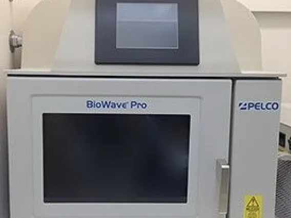
Pelco Biowave Pro Microwave Tissue Processor
Programmable microwave tissue processor, with continuous power settings from 100-750 Watts, load cooling to maintain consistent temperatures, with built-in fume extraction and vacuum.
The microwave is useful for:
- improved tissue fixation
- improved retention of fluorescence after fixation
- for more rapid tissue processing for immunocytochemistry
- electron microscopy
- decalcification.
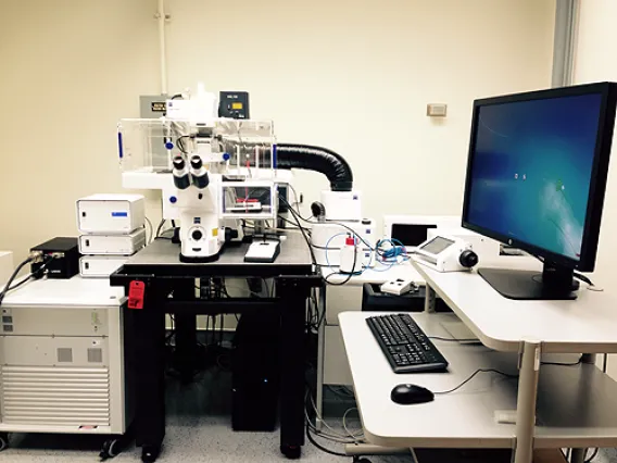
Zeiss Axiozoom 16 Fluorescent Stereo Microscope

Zeiss LSM880 NLO Upright Multiphoton/Confocal Microscope with Fast Airyscan
The Zeiss LSM 880 NLO upright multiphoton/confocal microscope includes a 34-channel detector for spectral information and additional non-descanned GAsP detectors. The system includes 7 visible spectrum laser lines (405, 458, 488, 514, 561, 633 nm), plus an additional software-tunable Spectra Physics Mai Tai HP DeepSee laser (690-1040 nm). The system includes software modules for controlled region-of-interest scanning, deconvolution, physiology experiments, performing FRAP & FCS and control of the motorized stage to allow stitching together images from multiple fields of view into one larger image (montage). The software also enables visiting multiple sites during a time lapse acquisition, and the instrument includes an environmental chamber suitable for live cell imaging at a variety of temperatures. Wet lab space is available to users of the facility in an adjacent room, including cell culture incubators and a biosafety hood.
NOTE: BSL2 samples must be discussed with the on-site core manager BEFORE they arrive in the facility. Live animals - invertebrates only!
The Fast Airyscan allows for resolution improvement up to 1.9x of typical confocal resolution and the fast mode sacrifices a bit of resolution for speed (useful for live cell imaging). The scanhead includes a 34-channel detector for spectral information.
|
Commonly used lenses on the Upright Zeiss |
Numerical |
Working |
type |
|---|---|---|---|
|
20x plan apo |
0.8 |
0.55 |
dry |
|
40x plan apo |
1.3 |
0.17 |
oil |
|
20x plan apo dippable |
1.0 |
2.4 |
water |
|
40x plan dippable |
0.8 |
2.1 |
water |

Zeiss LSM880 Inverted Confocal Microscope
The Zeiss LSM 880 inverted confocal microscope includes a 34-channel detector for spectral information, and 7 Laser lines (405, 458, 488, 514, 561, 594, 633nm). The system includes additional software modules for performing FRAP and control for the motorized stage to allow stitching together images from multiple fields of view into one larger image (montage), as well as visiting multiple sites during a time lapse acquisition. The instrument includes an environmental chamber suitable for live cell imaging at a variety of temperatures, humidity and CO2 capabilities. The facility has wet lab space, cell culture incubators, and a biosafety hood in an adjacent room.
NOTE: BSL2 samples must be discussed with the on-site core manager BEFORE they arrive in the facility.
Objective lenses (with matching DIC prisms) include:
- Plan-Apochromat 10x/0.45 (WD=2.0mm)
- Plan-Apochromat 20x/0.8 (WD=0.55mm)
- Plan-Apochromat 40x/1.3 Oil
- Plan-Apochromat 63x/1.4 Oil
- Plan-Apochromat 40x/0.95 dry CORR (not typically on the microscope, available upon request)
Zeiss SIM post-processing computer workstation
This workstation is only available to users already trained on the Zeiss Elyra S.1. First priority is given to users scheduled on the Zeiss Elyra S.1 for processing their data. The PC is a Hewlett-Packard Z6 workstation with an 8 core Intel Xeon CPU, 64GB of RAM, 12TB of data storage (RAID 10), running Windows 10 (64 bit) with a licensed version of the Zeiss ZEN software for data processing and FIJI (ImageJ).
Zeiss Elyra S.1 structured illumination microscope
The Zeiss Elyra S.1 uses the technique of structured illumination (SR-SIM) to achieve resolutions up to two times better than that of a confocal, deconvolution or standard fluorescence microscope. Resolutions in XY of 110nm and in Z of 300nm are possible (resolution is wavelength and sample-preparation dependent), allowing users to image a volume in the sample that is 1/8 that of previous techniques.
The good news is that this super-resolution technique is a not-too-difficult jump from the dyes and sample prep techniques used for a confocal or deconvolution microscope. The Elyra S.1 can image four fluorescent colors, roughly corresponding to DAPI, FITC/GFP, Rhodamine/RFP, and CY5 (see below). SR-SIM can image to depths of 10-15 microns into a sample. Note: SR-SIM does not work well (or at all) with dimly fluorescent samples or samples that photo-bleach.
The Elyra S.1 has the following microscope objectives:
- Plan-Neofluar 10x/0.30
- Plan-APO 40x/1.4 Oil
- Plan-Apochromat 63x/1.40 Oil
- Plan-Apochromat 63x/1.30 Oil/Water/Glycerine KORR
- Alpha Plan-APO 100x/1.46 Oil
The laser lines are 405, 488, 561, and 642nm. Imaging can be performed somewhat faster using a single multi-band cube, or with 4 sequential cubes to reduce spectral overlap:
- EF LBF 405/488/561/642
- EF BP 420-480 / BP 495-550 / LP 655
- EF BP 495-550/BP 570-620
- BP 420-480/LP 655
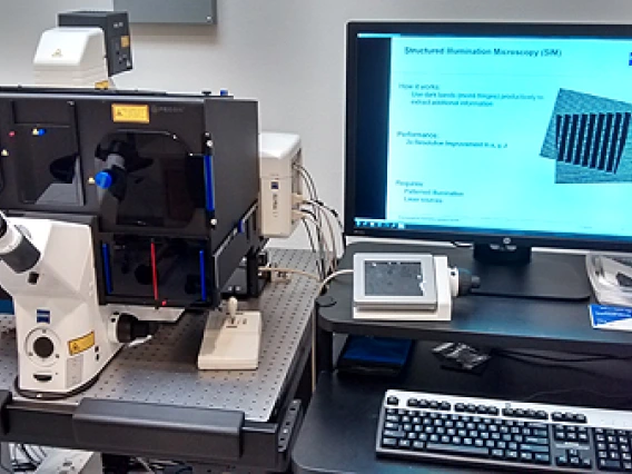
Image Analysis computer workstation
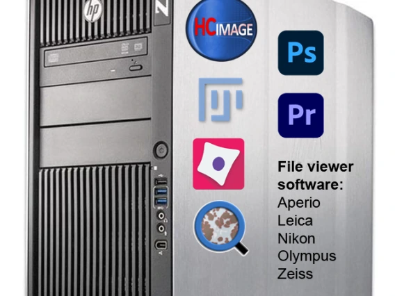
This Windows 10 workstation (HP Z820 with 8 Xeon cores and 128GB of RAM) is set up to run HCImage (Hamamatsu). HCImage can be configured with macros that allow the software to do semi-automated image analysis (using thresholding and segmentation) of image data from the DMI6000 microscope, or images from other sources. For calibrating images from other optical microscopes, Mr. Cromey has a stage micrometer that users can borrow. Numerical data from image analysis measurements can be exported as CSV files for further analysis (CSV files can be imported into MS Excel).
To use the workstation, contact Mr. Cromey first to discuss your image analysis needs. The workstation is free to use, however, if a macro needs to be built for your lab’s analysis, there may be several hours of billable labor involved to write it.
Other possible uses of this workstation include video editing and working with large files that might be too big to work with on standard lab computers/laptops. With the assistance of our IT team, the data directory on the Zeiss Axio Observer 7 with Apotome III and the Leica DMI6000 have been mapped so that they can be read on this workstation (read only access), allowing users to pull their data directly to this computer worktation if needed.
Software currently installed on this workstation (Jan 2023):
- HCImage 4.7 (initially purchased by the SWEHSC, P30 ES006694)
- ImageJ (FIJI) (open source image analysis software)
- Cell Profiler (open source image analysis software)
- Qu-Path (open source whole slide image analysis software)
- Free viewer software (Zeiss ZEN Blue, Nikon Elements viewer, Leica LAS X, Olympus viewer software, Aperio Scanscope)
- Adobe Creative Cloud (including Photoshop, Premier Pro) - (NOTE: you may need to have a license for these associated with your UA netID, if needed you can request access to Doug Cromey's license)
- Image data management resources:
- OMERO.insight (open source software for connecting to the campus OMERO server)
- CISCO AnyConnect (required for the UA VPN)
- Glencoe NGFF-converter (capable of batch transforming specific proprietary file formats into OME-TIF files, which may be required before they can be uploaded to the campus OMERO server)
Pagination
- Previous page
- Page 5
- Next page


